Conditions Treated
Cataract surgery remains the most frequently performed NHS surgical procedure in the UK with approximately 400,000 operations undertaken in England alone in a year.
What are cataracts?
Cataracts are when the lens in your eyes show signs of ageing and becomes cloudy and loose its transparency. When we are young, our lenses are usually clear like glass, allowing light to be focused on the retina and for us to see through them. As we age, they start to become frosted and begin to limit our vision.
What is a macula hole?
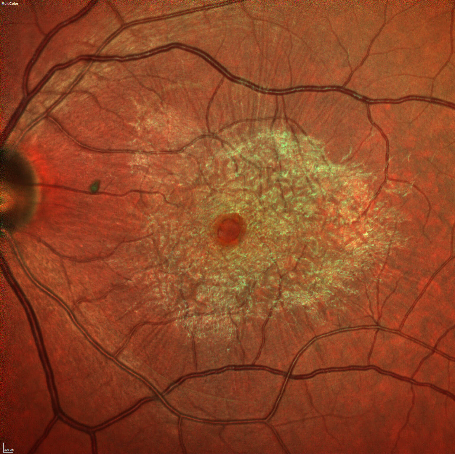
The retina is the layer of your eye that helps you see things. It is the equivalent of the film in a camera. The macula is the most sensitive part of your retina accounting for all of your fine central vision and colour perception. Occasionally, there can be a defect in the centre most point of your macula known as the macula hole.
What are epiretinal membranes?
Epiretinal membranes (also known as macular pucker or cellophane maculopathy) are fine membranes that grow on the surface of your retina. This membrane can distort and contort retinal structures resulting in vision being blurred and distorted.
What are floaters?
Floaters are small pieces of debris in the vitreous cavity (the jelly like substance within your eyes) of your eye. They are often described as either as small dark shapes that float across your vision and can look like spots, threads, squiggly lines, or even little cobwebs. Floaters move as your eyes move — so when you try to look at them directly, they seem to move away. When your eyes stop moving, floaters keep drifting across your vision.
You may notice floaters more when you look at something bright, like white paper or a blue sky.
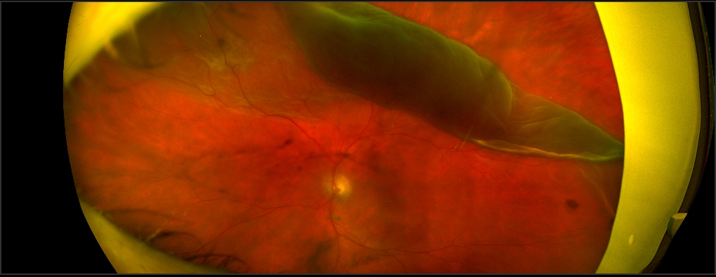
Retinal detachment is an ophthalmic emergency that requires an urgent assessment by an ophthalmologist and in most cases urgent surgery to prevent permanent sight loss. There are 3 types of retinal detachment: rhegmatogeneous (break related), tractional or exudative, each with different causes and treatment. We will discuss the rhegmatogeneous subtype which is the commonest cause of a retinal detachment.
How does Diabetes affect your eyes?
The retina is the light-sensitive layer of cells at the back of the eye that converts light into electrical signals. The signals are sent to the brain which turns them into the images you see.
The retina needs a constant supply of blood, which it receives through a network of tiny blood vessels. Over time, a persistently high blood sugar level can damage these blood vessels in 3 main stages:
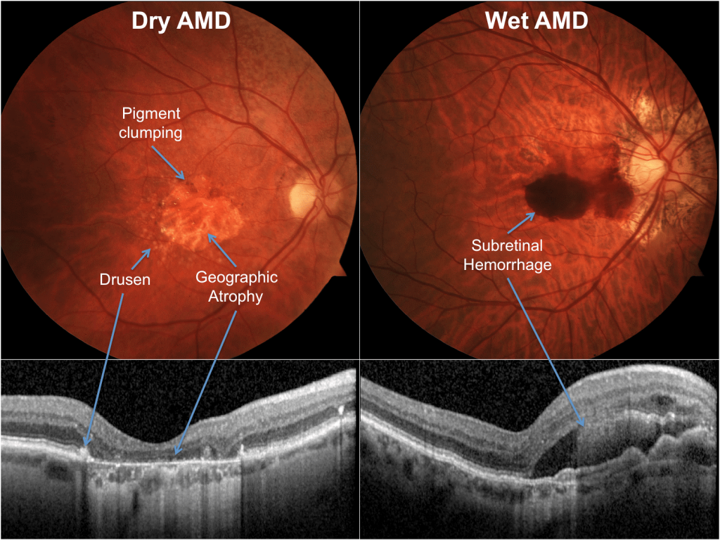
The advent of anti-vascular endothelial growth factors (anti VEGF) agents has revolutionised treatment for a number of ocular conditions including age related macular degeneration and cystoid macula oedema from diabetic maculopathy and retinal vein occlusions. Mr Jasani offers a detailed initial and follow up assessments, relevant investigation using state of the art imaging systems, and comprehensive treatment with retinal lasers and intravitreal injections for the above conditions.
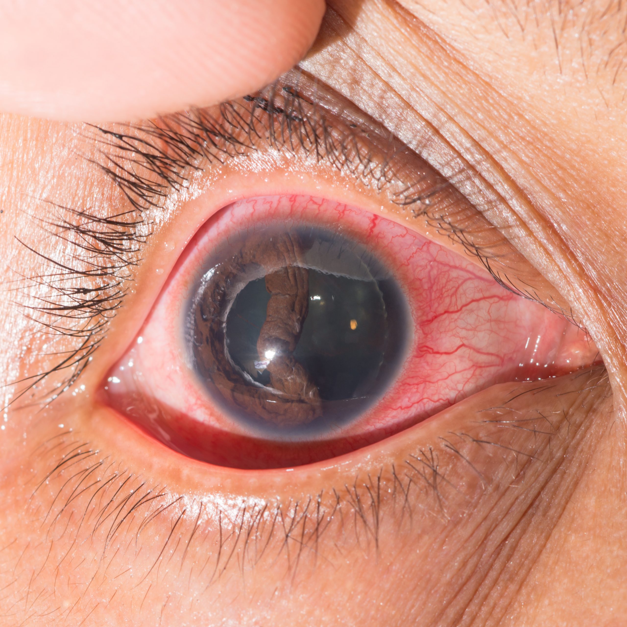
Unfortunately the eye is prone to injury and this can be separated into minor or severe trauma.
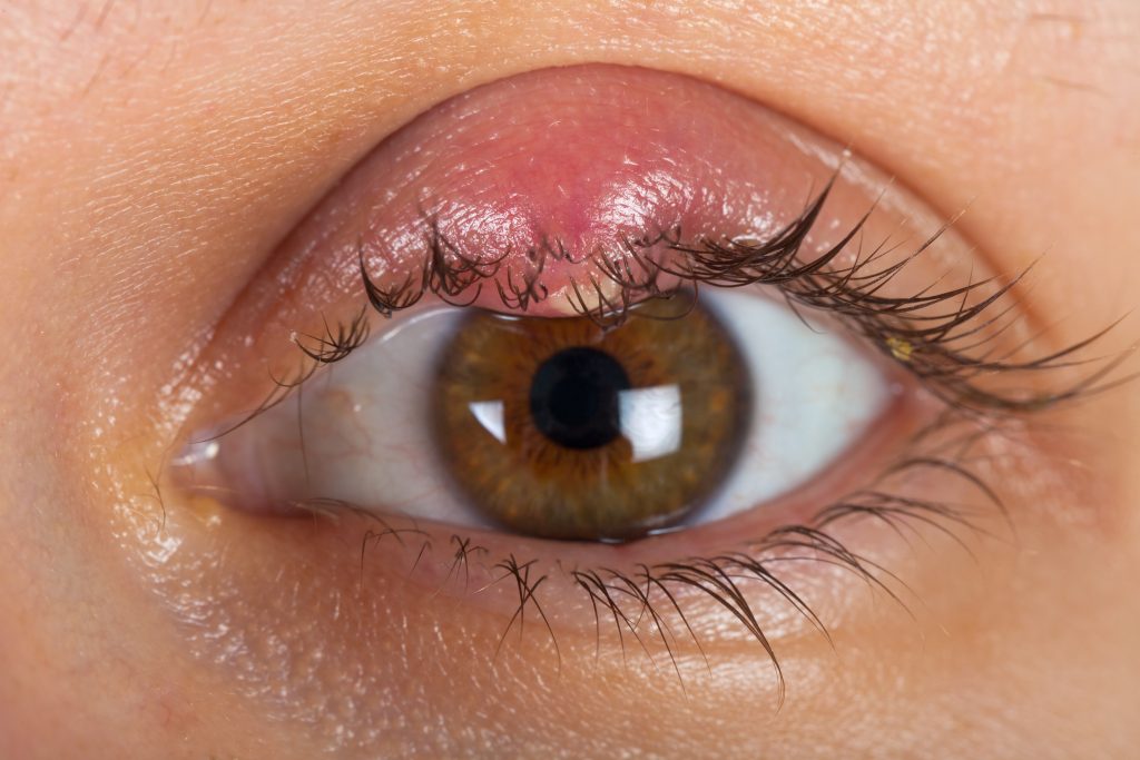
Chalazion and Stye
A chalazion is a cyst on the eyelid caused by a build up of secretions in the glands. A cyst is a round and usually painless swelling on the eyelid. Most often these will only shrink over many months and it is best to go forward to a small surgical procedure to drain the cyst. This can be performed under local anaesthetic and takes a few minutes.
A stye is a painful swelling of the eyelids caused by infection of the glands of the lid. It is usually treated by antibiotic ointment and hot spoon bathing.


